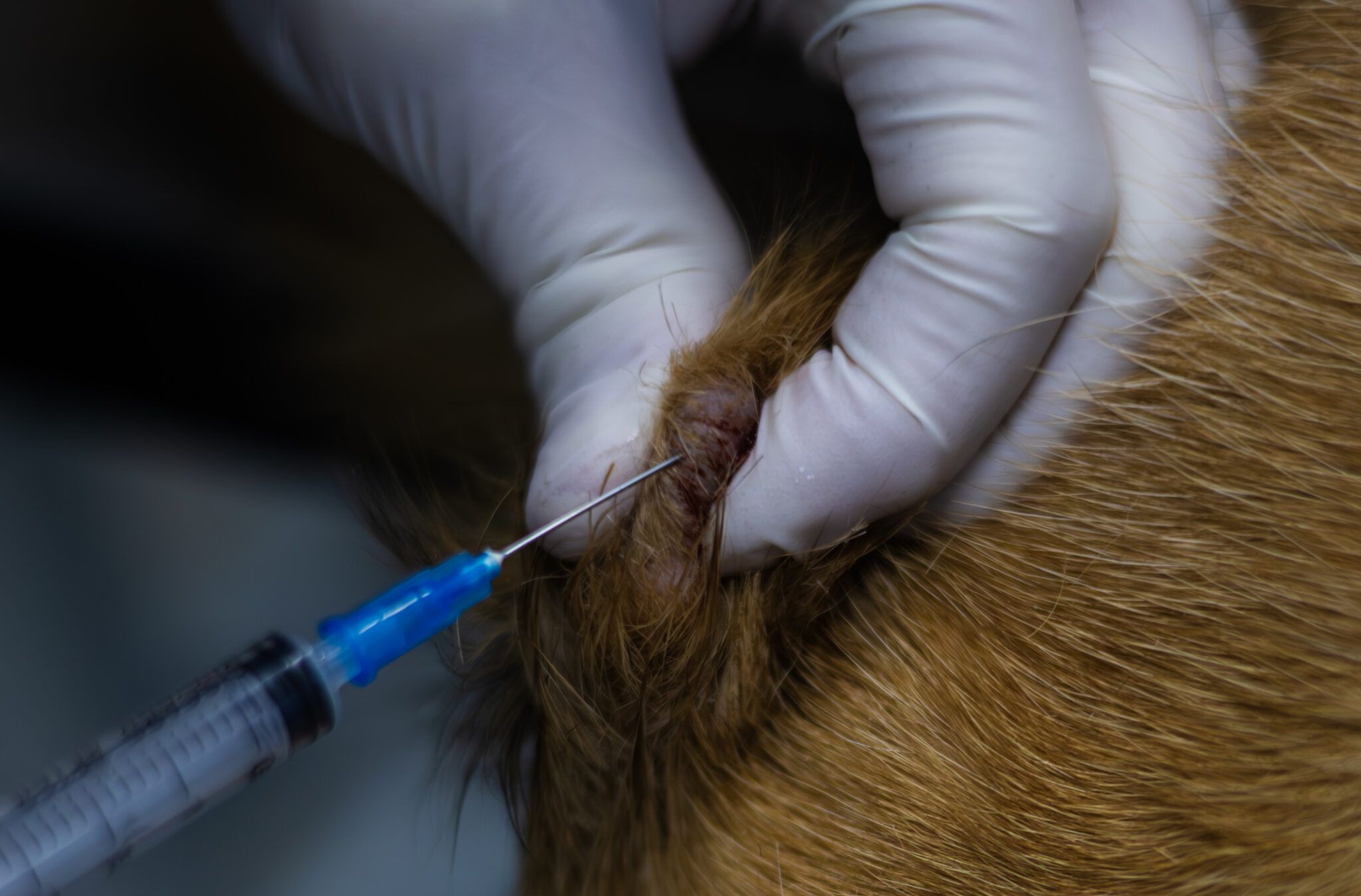
 You may opt to start determining if it’s a mast cell tumor or histiocytoma with a fine needle aspirate. This is simpler than surgery but gets cells to send to pathology for an initial diagnosis. Credit: Alexandr Lebedko | Getty Images
Dogs get bumps and growths on their skin. Not all are cancer, but interestingly, two physiologically very different bumps—mast cell tumors (cancerous) and histiocytomas (benign)—look so much alike that a new growth may cause you concern.
Because of this, if your dog develops a skin growth that looks like a mast cell tumor (MCT) or histiocytoma, it is best to have a fine needle aspirate done to look at the cells within the growth. This procedure is quick and easy; no sedation is required. Your veterinarian may look at the cells right at the clinic. If there are questions, slides will be sent for a veterinary pathologist’s evaluation.
Before that, let’s discuss the differences between the lookalike MCT and histiocytoma so you know what you may be up against.
A Histiocytoma Is Benign
Histiocytomas, also called “button tumors,” are small, raised, red lumps. They are almost always circular and most often found on the head, ears, or legs. The growth is smooth-surfaced and not itchy. They are almost always found on dogs under 3 years of age. Histiocytomas seem to appear more in short-haired dogs, although Flat Coat Retrievers have a high incidence.
If you are patient, a histiocytoma will usually fade away on its own, although it may take a couple of months. They do not metastasize.
In practice, clients with a dog with a histiocytoma ran about 50/50 as to watch and wait or remove — and all of them did fine.
MCTs Are Cancerous
Mast cell tumors are a different matter altogether. They tend to appear in older dogs. They may be subcutaneous or on the surface of the skin, and some will spread internally. Appearance can vary from benign-looking lumps to inflamed, irritated red growths or ulcers. If you rub on the lump, it may swell right before your eyes, with the skin getting red and warm. These growths are generally quite itchy. High-risk breeds include Boxers, Boston Terriers, Bulldogs, and Golden Retrievers, but any dog can develop mast cell tumors.
Mast cell tumors are graded according to size, location, and risk of metastasis. Prognosis is good for low-grade mast cell tumors, with worse outcomes for higher-grade masses. Because mast cells contain histamines, they may cause reactions such as vomiting, diarrhea, and stomach irritation. A fine needle aspirate can help with both diagnosis and grading.
Surgery is the mainstay of treatment. A low-grade mast cell tumor may be effectively cured by surgery and never recur. Unfortunately, dogs that have developed one are prone to recurrence, so vigilance is important going forward.
You may be surprised when you pick up your dog after surgery for a mast cell tumor. The growth was probably small, but the incision will be large. It is crucial with mast cell tumors to go as wide as possible to achieve clean margins — meaning no sign of cancer cells around the edges of the surgical site.
This is not something that can be assessed visually, which is why histopathology — sending samples to a lab — is essential. Follow-up radiation and/or chemotherapy may be recommended for aggressive mast cell tumors.
Because mast cell tumors and histiocytomas can look nearly identical to the naked eye, never assume a new skin growth is harmless. A quick fine needle aspirate gives you and your veterinarian the information needed to make the right call for your dog.